



Organismal Dynamics
Dinámica Nuclear en Biología Celular y del Desarrollo

Dr Peter Askjaer
Investigador/a Principal
Resumen
Para el desarrollo de un organismo se requiere una organización precisa de numerosos procesos biológicos. Concretamente, el control de la expresión génica y una correcta división celular son de suma importancia para todas las formas de vida, y llevan implícitas grandes interacciones entre proteínas y cromatina. En células eucariotas, el acceso al genoma está controlado por la envoltura nuclear (EN), que actúa de barrera selectiva de permeabilidad y organizadora de la cromatina.
La EN está constituida por tres componentes mayoritarios: las membranas nucleares, los complejos de poro nuclear (NPCs) y la lámina nuclear. Los NPCs están compuestos por múltiples copias de aproximadamente 30 nucleoporinas diferentes, y regulan el transporte entre el citoplasma y el núcleo. Mediante un análisis sistemático, hemos identificado varias de las nucleoporinas esenciales para la correcta segregación de los cromosomas y para la reconstitución de la EN tras la mitosis. Así, recientemente hemos descrito una interacción entre la nucleoporina NUP107/NPP-5 y la proteína del Spindle Assembly Checkpoint MAD1. Para nuestro estudio de nucleoporinas y otras proteínas de la EN combinamos análisis de microscopía óptica avanzada, RNAi de amplio espectro y ensayos bioquímicos.
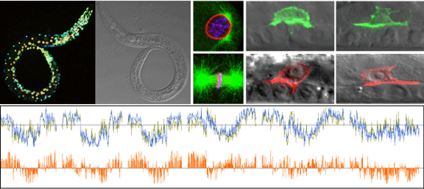
Por último, también aspiramos a aclarar la función biológica de la proteína kinasa conservada VRK-1. Mediante su fosforilación, las proteínas kinasas actúan como reguladoras de hasta un ~30% del proteoma humano. Entre los sustratos de VRK-1 se incluyen proteínas de la cromatina (por ejemplo, BAF, histonas) y factores de transcripción (por ejemplo, TP53, JUN). La pérdida de actividad de VRK-1 en mamíferos se asocia con esterilidad y fenotipos neurológicos, pero el mecanismo de las enfermedades no está claro. Hemos descrito defectos específicos subcelulares y en la organogénesis en mutantes C. elegans para vrk-1, y estamos analizando su posible relación con la fisiología humana.
Cinco publicaciones relevantes
Miembros del laboratorio y Colaboradores
Miembros del laboratorio
Investigador principal
Dr. Peter Askjaer (pask@upo.es)
Estudiantes de tesis
Celia Muñoz Jiménez
Raquel Romero Bueno
Patricia de la Cruz Ruiz
Estudiantes de Grado
Patricia Rider
Ana Blázquez Caraballo
Asistente de laboratorio
Cristina Ayuso García
Contacto
Despacho +34 954 348 396
Laboratorio +34 954 348 557
Ex-miembros del laboratorio
Dra. Elke PF Klerkx
Dra. Pilar Alarcón González
Dr. Eduardo Ródenas Martínez
Dra. María Jesús Rodríguez Palero
Dra. Cristina González Aguilera
Dra. Adela María Morales Martínez
Dra. Georgina Gómez Saldivar
Dra. Agnieszka Dobrzynska
Colaboradores
Dra Marta Artal-Sanz (CABD)
Dra Anita Fernandez (Uni Fairfield, CN, USA)
Dr Julien Dumont (JM, Paris, F) Dra Susan Gasser (FMI, Basel, CH)
Dr Peter Meister (Uni Bern, CH)
Dr Antonio Miranda-Vizuete (IBIS, Seville, ES) Dr Manuel J. Muñoz (CABD)
Plan Andaluz de Investigación, Desarrollo e Innovación (PAIDI)
Formamos parte del Grupo BIO295 – Mecanismos de Desarrollo Animal
FLP-Based Toolkit for Spatiotemporal Control of Gene Expression
Strains carrying single-copy transgenes expressing codon-optimised FLP
| Promoter | Cell Type | Strain | Reference | CGC |
| ckb-3 | Somatic gonad | BN854 | Fragoso-Luna et al | Yes |
| ceh-60 | Multiple | BN813 | Van de Walle et al | No |
| dat-1 | Dopaminergic neurons | BN617 | Muñoz-Jiménez et al | Yes |
| dpy-7 | Hypodermal | BN550 | Muñoz-Jiménez et al | Yes |
| eat-4 | Glutamatergic neurons | BN993 | Rossi et al | No |
| elt-2 | Intestine* | BN528 | Muñoz-Jiménez et al | Yes |
| gpa-14 | Neurons | BN1205 | Fragoso-Luna et al | No |
| hlh-8 | M lineage | BN502 | Muñoz-Jiménez et al | Yes |
| hlh-12 | Distal tip cell | BN1204 | Fragoso-Luna et al | Yes |
| hsp-16.41 | Ubiquitous | BN646 | Muñoz-Jiménez et al | Yes |
| lag-2 | Multiple | BN558 | Muñoz-Jiménez et al | Yes |
| lin-31 | Multiple | BN1023 | Fragoso-Luna et al | Yes |
| mec-7 | Mechanosensory neurons | BN498 | Muñoz-Jiménez et al | Yes |
| mex-5 | Germ line | BN711 | Macias-Leon & Askjaer | Yes |
| myo-2 | Pharyngeal muscle | BN543 | Muñoz-Jiménez et al | Yes |
| myo-3 | Body wall muscle | BN503 | Muñoz-Jiménez et al | Yes |
| nhr-82 | Seam cell lineage | BN455 | Muñoz-Jiménez et al | Yes |
| nhx-2 | Intestine | BN999 | Fragoso-Luna et al | Yes |
| rgef-1 | Pan-neuronal | BN507 | Muñoz-Jiménez et al | Yes |
| tph-1 | Serotonin-producing neurons | BN499 | Muñoz-Jiménez et al | Yes |
| UAS | Gal4-compatible | BN908 | Ayuso & Askjaer | Yes |
| unc-17 | Cholinergic neurons | BN1133 | Fragoso-Luna et al | No |
| unc-47 | GABAergic motor neurons | BN544 | Muñoz-Jiménez et al | Yes |
| unc-122 | Coelomocytes | BN1029 | Fragoso-Luna et al | Yes |
| ACEL-pes-10 | Anchor cell | AH5957 | Spiri et al | No |
* Note that recombination is also detected in the somatic gonad (see panels M-N below); this is not observed when the nhx-2 promoter is used to express FLP.
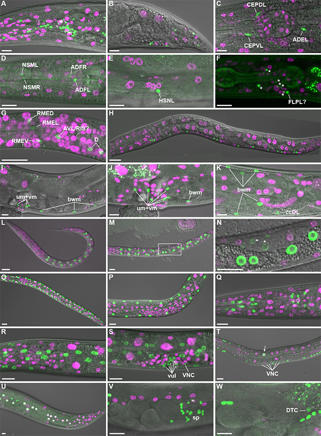
Figure 3 from Muñoz-Jiménez et al. Tissue-specific FLPase activity. Transgenic animals expressing FLP D5 in specific cell types and the dual color reporter were observed by live confocal microscopy. (A-B) Promoter of rgef-1 (Prgef-1); projection of eight confocal sections (A; adult head lateral view) or single confocal section (B; adult tail lateral view). (C) Pdat-1; projection of four confocal sections of head of L4 larva. Note that the green channel represents co-expressed GFP::HIS-58 and mNeonGreen in this strain (see Supplementary Figure 3). (D-E) Ptph-1; projection of three confocal sections (D; adult head lateral view) or a single confocal section (E; adult vulva lateral view). (F) Pmec-7; projection of seven confocal sections of head of L4 larva. To ease visualization of the weak GFP signal the corresponding DIC image was not included in the merge. Asterisks indicate six unidentified neurons, whereas a seventh neuron might be FLPL. (G-H) Punc-47; late L1; projection of four confocal sections (G; head lateral view) or stitch of single confocal sections (H). (I-K) Phlh-8; single confocal section (I; adult vulva lateral view) or max projections (J; vulva region; eight confocal sections, K; posterior gonad loop region; fifteen confocal sections). Examples of body wall muscle (bwm), coelomocyte (cc), uterine muscle (um) and vulval muscle (vm) cells are indicated. (L-N) Pelt-2; L1 (L; projection of seven confocal sections) and L2 larvae (M; projection of seven confocal sections, N; single confocal section corresponding to boxed area in M; note weak GFP expression in the gonad primordium [*]). (O-P) Pmyo-3; projection of seven confocal sections of L2 (O) and L4 (P) larvae. (Q) Pmyo-2; projection of twelve confocal sections of adult head. (R-T) Pdpy-7; projection of eight (R; head lateral view) or ten (S; vulva lateral view) confocal sections of L4 larva or single confocal section of L3 larva (T). Examples of vulva cells (vul) and ventral nerve cord neurons (VNC) are indicated. Arrow points to intestinal cell that expresses both markers. (U) Pnhr-82; projection of four confocal sections of tail of young adult. Note that white nuclei indicate simultaneous expression of mCh::HIS-58 and GFP::HIS-58. (V-W). Plag-2; projection of six confocal sections (V) or single confocal section (W) of central region of young adult. Spermatheca (sp) and distal tip cell (DTC) are indicated. All micrographs are oriented with anterior to the left and ventral down. Scale bar 10 µm.