



Organismal Dynamics
Nuclear Dynamics in Cell and Developmental Biology

Dr Peter Askjaer
Principal Investigator
Summary
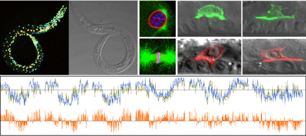
The nuclear envelope serves to protect and regulate the cell’s genetic material, the chromosomes, inside the cell nucleus. Interactions between chromosomes and proteins at the nuclear envelope are important for correct organization of chromosomes and gene transcription in a tissue-specific manner: although all cells in the body contain the same genes, they express different subsets of them and at different levels.
Our group at the Andalusian Centre for Developmental Biology studies the role of the nuclear envelope in these processes. We are interested in understanding the links between nuclear organization, development, diseases and aging. The nuclear envelope consists of the nuclear lamina, the outer and inner nuclear membranes and nuclear pore complexes that enable communication between the nucleus and the rest of the cell.
Five relevant publications
Lab members & Collaborators
Lab members
Group leader
Dr Peter Askjaer, pask@upo.es
PhD students
Raquel Romero Bueno
Patricia de la Cruz Ruiz
Paula Monterrubio Asensio
Research assistant
Cristina Ayuso García
Undergrad students
Laura Garzón
Sara Castaño Díaz
Nina Mellmann
Contacts
Office: +34 954 348 396
Lab: +34 954 348 557
Former lab members
Dr Elke PF Klerkx
Dr Pilar Alarcón González
Dr Eduardo Ródenas Martínez
Dr María Jesús Rodríguez Palero
Dr Cristina González Aguilera
Dr Adela María Morales Martínez
Dr Georgina Gómez Saldivar
Dr Agnieszka Dobrzynska
Dr Celia Muñoz Jiménez
Collaborators
Dr Marta Artal-Sanz (CABD)
Dr Julien Dumont (JM, Paris, F)
Dr Anita Fernandez (Uni Fairfield, CN, USA)
Dr Susan Gasser (FMI, Basel, CH)
Dr Peter Meister (Uni Bern, CH)
Dr Antonio Miranda-Vizuete (IBIS, Seville, ES)
Dr Manuel J. Muñoz (CABD)
Plan Andaluz de Investigación, Desarrollo e Innovación (PAIDI)
We form part of the group BIO295 – Mechanisms of Animal Development
FLP-Based Toolkit for Spatiotemporal Control of Gene Expression
Strains carrying single-copy transgenes expressing codon-optimised FLP
| Promoter | Cell Type | Strain | Reference | CGC |
| ckb-3 | Somatic gonad | BN854 | Fragoso-Luna et al | Yes |
| ceh-60 | Multiple | BN813 | Van de Walle et al | No |
| dat-1 | Dopaminergic neurons | BN617 | Muñoz-Jiménez et al | Yes |
| dpy-7 | Hypodermal | BN550 | Muñoz-Jiménez et al | Yes |
| eat-4 | Glutamatergic neurons | BN993 | Rossi et al | No |
| elt-2 | Intestine* | BN528 | Muñoz-Jiménez et al | Yes |
| gpa-14 | Neurons | BN1205 | Fragoso-Luna et al | No |
| hlh-8 | M lineage | BN502 | Muñoz-Jiménez et al | Yes |
| hlh-12 | Distal tip cell | BN1204 | Fragoso-Luna et al | Yes |
| hsp-16.41 | Ubiquitous | BN646 | Muñoz-Jiménez et al | Yes |
| lag-2 | Multiple | BN558 | Muñoz-Jiménez et al | Yes |
| lin-31 | Multiple | BN1023 | Fragoso-Luna et al | Yes |
| mec-7 | Mechanosensory neurons | BN498 | Muñoz-Jiménez et al | Yes |
| mex-5 | Germ line | BN711 | Macias-Leon & Askjaer | Yes |
| myo-2 | Pharyngeal muscle | BN543 | Muñoz-Jiménez et al | Yes |
| myo-3 | Body wall muscle | BN503 | Muñoz-Jiménez et al | Yes |
| nhr-82 | Seam cell lineage | BN455 | Muñoz-Jiménez et al | Yes |
| nhx-2 | Intestine | BN999 | Fragoso-Luna et al | Yes |
| rgef-1 | Pan-neuronal | BN507 | Muñoz-Jiménez et al | Yes |
| tph-1 | Serotonin-producing neurons | BN499 | Muñoz-Jiménez et al | Yes |
| UAS | Gal4-compatible | BN908 | Ayuso & Askjaer | Yes |
| unc-17 | Cholinergic neurons | BN1133 | Fragoso-Luna et al | No |
| unc-47 | GABAergic motor neurons | BN544 | Muñoz-Jiménez et al | Yes |
| unc-122 | Coelomocytes | BN1029 | Fragoso-Luna et al | Yes |
| ACEL-pes-10 | Anchor cell | AH5957 | Spiri et al | No |
* Note that recombination is also detected in the somatic gonad (see panels M-N below); this is not observed when the nhx-2 promoter is used to express FLP.
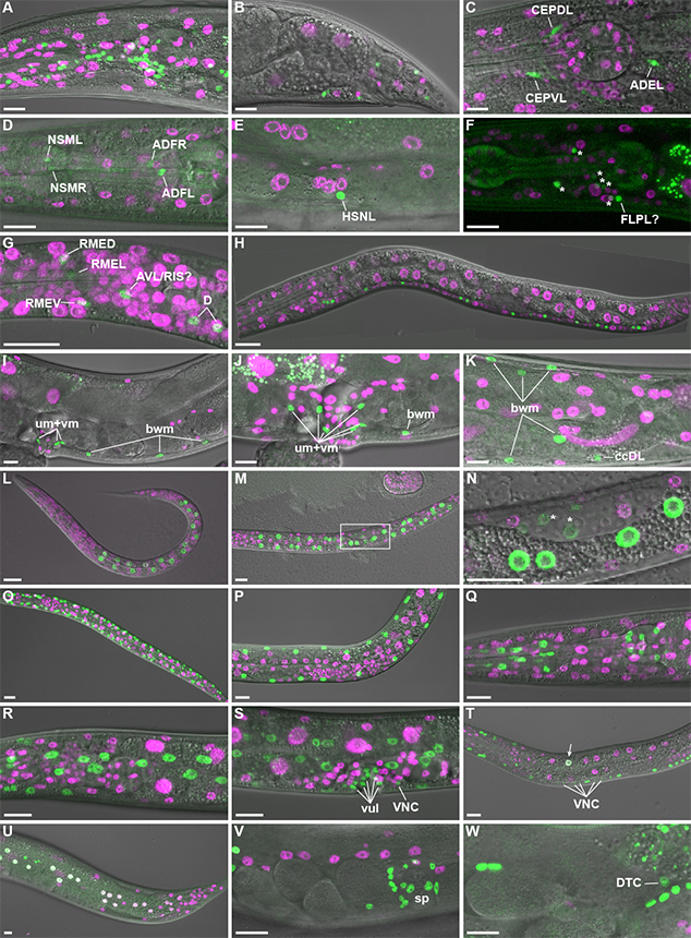
Figure 3 from Muñoz-Jiménez et al. Tissue-specific FLPase activity. Transgenic animals expressing FLP D5 in specific cell types and the dual color reporter were observed by live confocal microscopy. (A-B) Promoter of rgef-1 (Prgef-1); projection of eight confocal sections (A; adult head lateral view) or single confocal section (B; adult tail lateral view). (C) Pdat-1; projection of four confocal sections of head of L4 larva. Note that the green channel represents co-expressed GFP::HIS-58 and mNeonGreen in this strain (see Supplementary Figure 3). (D-E) Ptph-1; projection of three confocal sections (D; adult head lateral view) or a single confocal section (E; adult vulva lateral view). (F) Pmec-7; projection of seven confocal sections of head of L4 larva. To ease visualization of the weak GFP signal the corresponding DIC image was not included in the merge. Asterisks indicate six unidentified neurons, whereas a seventh neuron might be FLPL. (G-H) Punc-47; late L1; projection of four confocal sections (G; head lateral view) or stitch of single confocal sections (H). (I-K) Phlh-8; single confocal section (I; adult vulva lateral view) or max projections (J; vulva region; eight confocal sections, K; posterior gonad loop region; fifteen confocal sections). Examples of body wall muscle (bwm), coelomocyte (cc), uterine muscle (um) and vulval muscle (vm) cells are indicated. (L-N) Pelt-2; L1 (L; projection of seven confocal sections) and L2 larvae (M; projection of seven confocal sections, N; single confocal section corresponding to boxed area in M; note weak GFP expression in the gonad primordium [*]). (O-P) Pmyo-3; projection of seven confocal sections of L2 (O) and L4 (P) larvae. (Q) Pmyo-2; projection of twelve confocal sections of adult head. (R-T) Pdpy-7; projection of eight (R; head lateral view) or ten (S; vulva lateral view) confocal sections of L4 larva or single confocal section of L3 larva (T). Examples of vulva cells (vul) and ventral nerve cord neurons (VNC) are indicated. Arrow points to intestinal cell that expresses both markers. (U) Pnhr-82; projection of four confocal sections of tail of young adult. Note that white nuclei indicate simultaneous expression of mCh::HIS-58 and GFP::HIS-58. (V-W). Plag-2; projection of six confocal sections (V) or single confocal section (W) of central region of young adult. Spermatheca (sp) and distal tip cell (DTC) are indicated. All micrographs are oriented with anterior to the left and ventral down. Scale bar 10 µm.