



Genome Dynamics
Cell Potency Transitions

Dr. Thomas Spruce
Researcher
Summary
Cell potency describes a cells capacity to differentiate into other cell types, with higher potency indicating greater plasticity. Generally, cell potency decreases as embryo development proceeds however this is not the case at the beginning of the process when, following fertilisation, a highly differentiated egg cell gives rise to ball of totipotent cells capable of forming all the lineages of the embryo. In mammals the emergence of totipotency is intimately linked both to the maternal zygotic transition, in which awakening of the transcriptionally inactive zygotic genome is coupled with degradation of maternally inherited transcripts, and a dramatic epigenetic reprogramming of the embryo. A totipotent state is then retained through early cleavages until the morula stage when the embryo segregates into the placenta forming trophectoderm (TE) and foetus forming inner cell mass (ICM). A further decrease in potency is seen at the blastocyst stage when the inner cell mass splits into the primitive endoderm, which will give rise to the visceral endoderm and yolk sack and the pluripotent epiblast which will give rise to the foetus itself. Embryo implantation is accompanied by a further change in potency with the epiblast transitioning from a naïve state, away from differentiation to primed one, competent to respond to differentiation cues. Both naïve and primed stem cells can be derived from the embryo at pre and post implantation stages respectively and kept in culture. Furthermore, studies during the last decade have found that cultured embryonic stem cells (ESCs) regularly transition through a state that is similar to the 2-cell embryo and that this can be promoted through the use of various combinations of small molecules.
The overriding objective of my research is to uncover the mechanisms underlying mammalian totipotency and pluripotency acquisition and loss both in the embryo and in cell culture systems. To begin with we are focussing on the role of the Hippo signalling pathway in these processes which has been implicated in zygotic genome activation, trophectoderm formation and epiblast/primitive endoderm segregation. For this we are making use of a wide variety of experimental approaches including CRISPR mediated genome editing, high throughput sequencing, embryo micromanipulation and confocal imaging.
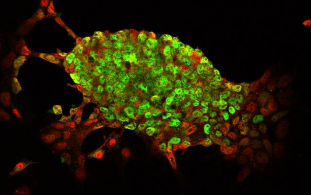
Cells at the edge of ES cell colonies have reduced expression of the pluripotency marker Nanog (green) and increased nuclear localisation of Yap1 (red).
Relevant literature
Lab members & collaborators
Lab members
Associated Investigator
Dr. Thomas Spruce, tspr1@upo.es
Masters student
Marta Escobar
Collaborators
Dr. Bárbara Pernaute (CABD, Sevilla)